Proceedings · Session S-258 · filed September 30, 2026
Translational ScienceSession paper
0.55 mm Piezoelectric Microprobe Cuts IV-OCT Catheter Size Nearly Fourfold
A 0.55 mm piezoelectric IV-OCT probe from Nanjing researchers cut angular deviation to 1° at 50 rev/s and reached the middle cerebral artery in a human vascular model.
By Amara Osei3 min read602 words
Summary
- The probe measures 0.55 mm in diameter, optimal for 2 mm vessels, versus ~2 mm conventional IV-OCT catheters suited to 10 mm vessels.
- Bench tests in a curved vessel phantom showed 1° angular deviation at 50 revolutions per second, compared with 9° for traditional proximal catheters.
- The probe traversed a full-scale human vascular model to the middle cerebral artery and its plaque images agreed with histology on rupture sites and collagen-rich regions.
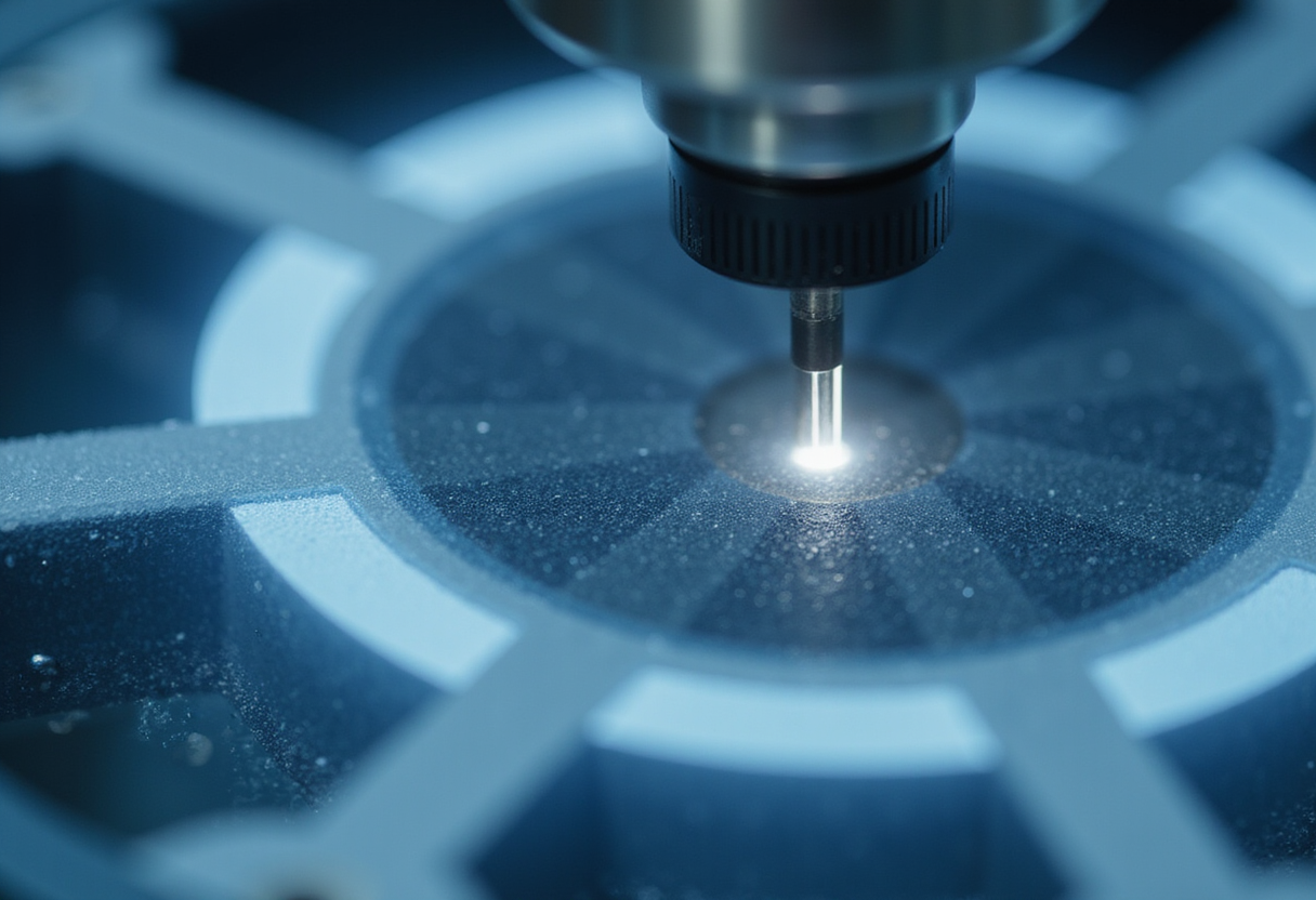
Researchers at Nanjing University of Aeronautics and Astronautics and Nanjing University Medical School have built an intravascular optical coherence tomography (IV-OCT) probe with a diameter of 0.55 mm — roughly a quarter the size of the ~2 mm catheters used clinically today — and demonstrated it in ex vivo pig vessels, human plaques and a full-scale human vascular model. The work, led by Dawei Wu and Rui Liu with first author Boquan Wang, appears in Biomedical Optics Express.
The size reduction matters because conventional IV-OCT catheters perform optimally in vessels around 10 mm in diameter — medium and large arteries. Electromagnetic motors and their wiring set the floor on how small these probes can get. Atherosclerotic disease, responsible for roughly three-quarters of cardiovascular deaths worldwide, also afflicts far smaller vessels, including the cerebrovascular network, which current catheters cannot reach.
A different drive mechanism
The two dominant catheter designs each carry a well-documented trade-off. Proximal systems, which generate rotation outside the body, suffer friction-induced rotational distortion in small, tortuous vessels. Distal designs place a motor inside the catheter but lose part of the 360° view to wire artefacts. The Nanjing group sidestepped both problems by replacing the electromagnetic motor with a piezoelectric drive.
The microprobe runs on a single-phase AC circuit that drives a piezoelectric crystal to vibrate a glass tube longitudinally. A 10° groove in the glass converts that longitudinal vibration into torsional vibration, producing elliptical motion of the lens — mechanically analogous to a crank and slider. Applying AC voltage makes the crystal expand and contract, rotating the lens and generating the optical scan. Because rotation needs only single-phase voltage, the wiring stays minimal, which is what shrinks the overall diameter.
Measured performance
The team first tested rotational fidelity by imaging metal tubes arranged around a small, curved vessel. The probe scanned at 50 revolutions per second while holding angular deviation to 1°. Traditional proximal IV-OCT catheters show 9° of deviation under comparable conditions, according to the authors' reference. The same test confirmed the full 360° field of view.
Navigation and imaging followed in leaf microveins, a vascular stent and ex vivo pig vessels. In a milestone for cerebrovascular applications, the probe traversed a full-scale human vascular model and reached the middle cerebral artery — an anatomical target no commercial IV-OCT catheter can image today.
To validate diagnostic capability, the researchers compared probe-acquired OCT images of human plaques against histological analyses. The two methods agreed well on plaque rupture sites and collagen-rich regions.
What is measured, what is projected
The 1° angular deviation, 50 revolutions per second, 0.55 mm diameter and 360° field of view are bench and ex vivo measurements. The clinical value — plaque pathology assessment and stent planning in the heart–brain system's smaller, more curved vessels — remains a projection that will require further development and testing. The human vascular model traversal, while encouraging, is a phantom result, not an in vivo one, and the paper does not report imaging resolution figures in this account.
"Although the probe would need more development and testing to be used clinically, intravascular OCT could one day give physicians a much closer look at what is happening inside the small arteries of the brain," Wu said in a press statement.
For R&D managers in imaging and medtech portfolios, the result signals that piezoelectric drive mechanisms can break the electromagnetic-motor size barrier that has constrained intravascular OCT for years. The Nanjing team's next hurdles are the translation from ex vivo and phantom studies to clinical validation in living patients.
via ahajournals.org (Original)
Filed under
- medical-imaging
- piezoelectrics
- cardiovascular
- optical-coherence-tomography
- medical-devices
More from Amara Osei
References
- MIT Team Grows Steerable Blood Vessels Using Magnetic Forces
- Light-Sheet Microscope Tracks Zebrafish Seizures in 3D at Four Volumes per Second
- Siemens Healthineers launches Accela linac for 60-second SBRT
- Monte Carlo workflow quantifies motion damage in proton minibeam therapy
- NASA Bets on Compact Lab Hardware to Reshape Research Workflow