Proceedings · Session S-119 · filed October 10, 2026
Lab Technology & MethodsSession paper
MIT's portable 3D breast ultrasound lifts target detection to 79.7%
MIT's 3D PURE portable ultrasound detected 79.7% of phantom microtargets versus 60.7% for handheld 2D, with a 10% resolution gain from adaptive beamforming.
By Sophie Lindqvist4 min read845 words
Summary
- 3D PURE detected 79.7% of sub-millimetre phantom targets versus 60.7% for a conventional 2D handheld system, with detection efficiency of 14.02% vs 9.58% targets per minute.
- The 128-element transducer's corner-gap offset geometry suppresses peak crosstalk by 3.73 dB and supports transmit voltages up to 24 V.
- The LACR adaptive beamformer delivers up to a 10% resolution improvement by compensating for heterogeneous breast tissue sound speeds.
- Seven novice users achieved 94.10% mean probe-placement overlap on frontal projections and 87.42% on side projections using the MyFUS interface.
- Interval cancers arising between screenings account for 20–30% of all breast cancer cases; the work appears in Nature Communications.
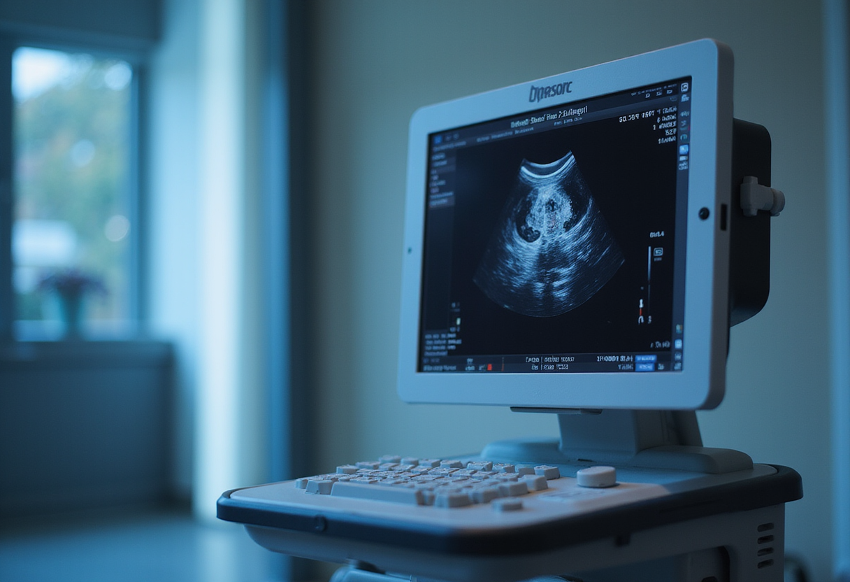
A portable ultrasound system developed at MIT detected 79.7% of sub-millimetre targets embedded in a breast phantom, against 60.7% for a conventional 2D handheld system, in an in vitro study with ten participants. The device, described in Nature Communications, is intended to catch fast-growing "interval cancers" that arise between routine screening exams — a category that accounts for 20% to 30% of all breast cancer cases.
The 3D portable ultrasound system for real-time examination (3D PURE) generates high-resolution 3D images of breast tissue and supplements mammography, whose sensitivity drops in dense breast tissue. Principal investigator Canan Dagdeviren's team at the MIT Media Lab positions the system as a tool for frequent longitudinal imaging performed not only by nurses, primary care physicians or gynaecologists, but potentially by patients themselves.
What does the second-generation hardware change?
This prototype is the successor to an earlier MIT design, and the improvements are specific and measurable. The team built a 128-element box-shaped 2D transducer array with a corner-gap offset geometry that suppresses peak crosstalk by 3.73 dB at the corner-most element. That suppression prevents amplifier saturation and supports transmit voltages up to 24 V. The array sits in a compact multilayer electronics stack containing preamplifiers, transmit electronics and a custom chirp data acquisition system.
A conductive backing layer — a toluene-diluted polyurethane matrix loaded with a high percentage of tungsten and zirconia filler — surrounds the elements continuously. According to the researchers, this improves acoustic attenuation, provides electromagnetic shielding and dampens reverberations.
"The addition of a backing layer to the ultrasound transducer is a key advantage to the system," said co-lead author Md Osman Goni Nayeem. "It provides both better directionality of ultrasound waves and better bandwidth, which improves the resolution and quality of the resulting images."
The team also integrated layered aberration-correction reconstruction (LACR), an adaptive 3D beamformer that compensates for the heterogeneous speed of sound in breast tissue — glandular and fibrous tissue plus a superficial fat layer. The researchers believe this is the first wide-angle 3D ultrasound imaging system to implement aberration-correction beamforming.
"What we are trying to do is predict the speed-of-sound properties of the tissue being imaged, and then use that to reconstruct the image more accurately. We see up to a 10% improvement for the resolution just by applying this [beamforming] technique," said co-lead author Shrihari Viswanath in a press statement.
How usable is it for non-experts?
The system's operator dependence question is central to its deployment case. A visual interface called "Mirror my First UltraSound" (MyFUS) guides users to reposition the probe at the same anatomical location across repeated scans, and a wide field-of-view limits the number of scans needed to cover the breast.
The team tested this claim directly. Seven volunteers with no prior ultrasound experience received initial instruction from an ultrasound technician, then repeatedly positioned the probe at specific locations on their breasts. They achieved a mean overlap of 94.10% on frontal projections and 87.42% on side projections — evidence, the researchers argue, that novices can produce reproducible longitudinal monitoring data.
In the phantom study, nine of the ten participants detected more microtargets with 3D PURE than with the handheld 2D system, and detection efficiency reached 14.02% of available targets per minute versus 9.58% for the conventional device.
What did the in vivo validation show?
Working with radiologists, the team assessed a range of breast anomalies in living subjects. The radiologists verified that 3D PURE could accurately visualize calcifications, cysts, implants, fibrous tissues and solid masses within a large volumetric field-of-view. The researchers also independently validated the system's accuracy when imaging dense fibrous breast tissue and rib structures. The published comparison images show conventional 2D views of a cyst, a breast implant, dense fibrous tissue and a solid mass alongside the corresponding 3D PURE reconstructions.
The validation scope has limits that R&D and clinical readers should weigh. The phantom study involved ten participants; the usability study, seven; the in vivo assessment was a radiologist review of anomaly visualization rather than a diagnostic accuracy trial against a clinical reference standard. The 10% resolution improvement from LACR is a measured figure; broader claims about interval-cancer detection remain projections until trial data exist.
Who could use it next?
The researchers' stated roadmap targets decentralization. "The high operator dependence of conventional ultrasound is a barrier to its use in decentralized or home-monitoring settings," they write. "The MyFUS vision interface transforms longitudinal monitoring from an expert-driven procedure to a self-guided, reproducible process that can be used by novice users. This reproducibility is vital for longitudinal monitoring in limited clinical settings, to regularly track the growth of an anomaly or the response of a tumour to therapies without visiting clinics."
The team's next step is an interface for mobile phones or tablets, aimed at patients in economically constrained countries without adequate breast cancer screening capacity and at regions facing shortages of trained ultrasound technicians.
via mit.edu (Original)
Filed under
- 3d-ultrasound
- breast-cancer-screening
- medical-imaging
- point-of-care-diagnostics
- transducer-technology
More from Sophie Lindqvist
Show full bio
Correspondent covering business strategy at Hypothesis Wire.
149 articles
References
- Siemens Healthineers launches Accela linac for 60-second SBRT
- Leica Ships Laser Cassette Printer Rated at 2.5 Seconds Per Cassette
- Ultrasound drug delivery peaks at 15–23 nm, UVA team finds
- Rare Earth Nanoparticles Track HER2, EGFR, HER3 for 16 Minutes Straight
- Siemens Healthineers Puts €10m Into Lab Equipment R&D Centre in Ireland