Proceedings · Session S-565 · filed November 6, 2026
Translational ScienceSession paper
Study Locates the Genomic Sites Where PARP Inhibitors Kill Cancer Cells
NYU Langone researchers show PARP inhibitor cytotoxicity depends on where replication fails — pericentromeric regions — not just how much DNA damage accumulates.
By Priya Raman4 min read734 words
Summary
- Study published in Nature Cell Biology: 'Positional replication fork instability drives PARP inhibitor cytotoxicity', led by NYU Langone Health's Perlmutter Cancer Center.
- CRISPR screens in breast and small cell lung cancer models identified ESCO2 as a strong determinant of PARP inhibitor sensitivity.
- Partial ESCO2 loss increased sensitivity in colorectal, ovarian and lung cancer cells while homologous recombination remained intact.
- Damage concentrated in repetitive pericentromeric regions; the protective response involved PARP1, TIMELESS and DDX11.
- Work was done in cancer cell models only; clinical validation in patient-derived cells is underway with clinician partners.
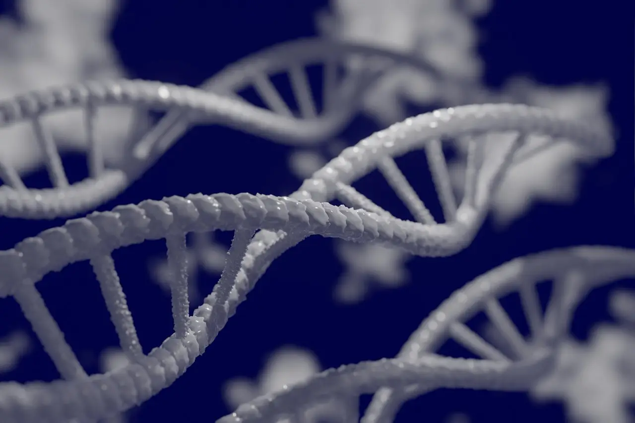
Partial loss of a single chromosome-cohesion protein, ESCO2, made breast, colorectal, ovarian and lung cancer cells more sensitive to PARP inhibitors — without disabling the homologous recombination repair pathway that clinicians usually require before prescribing the drugs. That result, published in Nature Cell Biology under the title "Positional replication fork instability drives PARP inhibitor cytotoxicity," reframes the question of why these therapies work: not how much DNA damage a cell accumulates, but where on its chromosomes that damage lands.
The study was led by researchers at NYU Langone Health's Perlmutter Cancer Center. Eli Rothenberg, PhD, professor of biochemistry and molecular pharmacology at NYU Grossman School of Medicine, and Dipika Gupta, PhD, a postdoctoral scholar in his laboratory, served as co-corresponding authors.
The work carries direct implications for R&D portfolios. PARP inhibitors are approved chiefly for BRCA-mutated cancers, where homologous recombination is already impaired. If sensitivity can instead be driven by replication stress at specific genomic positions, the eligible patient population for this drug class could widen considerably — provided the findings survive preclinical and clinical testing.
How the researchers found the signal
The team started with CRISPR screens in breast and small cell lung cancer models. The screens flagged ESCO2, a protein that helps hold newly copied chromosomes together, as a strong determinant of PARP inhibitor sensitivity. Even partial depletion of ESCO2 increased drug sensitivity in colorectal, ovarian and lung cancer cells, and the ESCO2-depleted cells retained the tested homologous recombination response.
To explain that sensitivity, the researchers used quantitative single-molecule imaging, an ultrasensitive microscopy method developed in Rothenberg's laboratory. The technique distinguishes subtypes of replication-associated damage sites that other methods cannot readily separate, according to Rothenberg.
"The imaging enabled us to move beyond measuring damage across an entire cell," Gupta said. "We could detect subtle replication problems, distinguish the sites involved, and identify the protective response that the cells relied on."
What did the imaging show?
Reducing ESCO2 increased G-quadruplexes — folded DNA structures that can obstruct copying. The increase was not distributed evenly across the genome. It concentrated at a small subset of replication sites, particularly in repetitive pericentromeric regions adjacent to centromeres.
That protective response depended on PARP1 activity and involved two additional proteins: TIMELESS, which helps protect DNA replication, and DDX11, an enzyme that can unwind G4 structures. When the researchers blocked PARP, the response failed in ESCO2-depleted cells. G4s accumulated further, DNA synthesis declined and damage increased near centromeres — a mechanism that helps explain the cells' sensitivity to the drugs.
"We often think of DNA damage in terms of quantity: More damage means a greater chance of killing a cancer cell," Rothenberg said. The findings point instead to location as a decisive factor in treatment response.
What are the limits of the data?
Several caveats deserve attention from anyone weighing translational claims. The study was conducted in cancer cell models, not in animals or patients. The authors themselves state that further preclinical and clinical research is needed to establish whether these replication-stress features can guide treatment decisions. And the proposal that other DNA-damaging treatments may work partly because they preferentially hit these sensitive genomic sites remains, for now, an untested hypothesis — the paper notes that whether it holds in other genetic contexts and with other treatments must be examined directly.
The measured results to date are these: ESCO2 loss confers PARP inhibitor sensitivity across at least four cancer types in vitro; the damage concentrates in pericentromeric regions; and the cytotoxic mechanism runs through PARP1, TIMELESS and DDX11. Everything beyond that — biomarker development, patient stratification, combination strategies — is projection.
Still, the translational path has begun. Rothenberg is working with clinicians to apply the laboratory's imaging approaches to patient-derived cancer cells, with the goal of identifying patterns of replication stress and DNA damage that could help predict who responds to treatment. The authors also argue the results lay a foundation for biomarkers of cancer drug sensitivity.
"Our work reveals a genomic Achilles' heel — a small subset of sites where replication problems can have disproportionately severe consequences," Rothenberg said. "Identifying those sites helps explain what cancer cells depend on to survive and why a particular treatment can be so effective."
For assay developers and oncology pipeline managers, the next concrete checkpoint will be whether pericentromeric replication-stress signatures measured in patient-derived cells correlate with clinical response to PARP inhibitors.
via nature.com (Original)
Filed under
- parp-inhibitors
- cancer-research
- dna-replication-stress
- crispr-screening
- single-molecule-imaging
More from Priya Raman
Show full bio
Staff writer covering money, funding and market shifts around research and development at Hypothesis Wire.
247 articles
References
- Merck Pays $400M Upfront for Preclinical KRAS G12D Glue in $2.13B Deal
- Recursion Targets H2 2027 Pivotal Trial After REC-4881 Cuts FAP Polyps 47%
- Roche Taps Defand in $1B Molecular Glue Discovery Pact
- Cancer Cell publishes sac-TMT plus osimertinib translational data
- Elephas Biosciences and TES Pharma Partner on Lung Cancer Translational Research