Proceedings · Session S-516 · filed November 4, 2026
Translational ScienceSession paper
One-Million-Cell Glioblastoma Atlas Shows Tumors Follow Rules, Not Chaos
A Nature study maps over one million glioblastoma cells from 12 patients and finds a conserved, predictable cell trajectory, with a free atlas and a companion paper on gene regulators.
By Priya Raman4 min read801 words
Summary
- Researchers mapped over 1 million cells across glioblastoma tumors from 12 patients, published in Nature.
- Cancer cells follow a conserved trajectory: developmental-like edge states transition to injury-response and then hypoxic core states across all patients.
- Average glioblastoma survival is 12 to 18 months; the atlas and companion regulatory study are both in Nature.
- The GBM-Space atlas is freely accessible online for the research community.
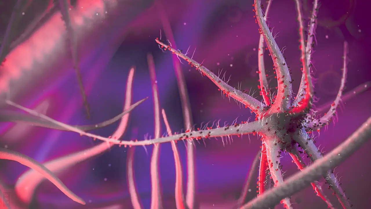
Researchers have mapped more than one million cells from glioblastoma tumors and found that the cancer organizes itself into the same structural pattern across patients, overturning the long-held view that the disease is molecularly chaotic. The study, published in Nature under the title "A spatiotemporal cancer cell trajectory underlies glioblastoma heterogeneity," analyzed tumor samples from 12 glioblastoma patients and was reported by scientists at the Wellcome Sanger Institute, the European Molecular Biology Laboratory (EMBL), the German Cancer Research Center (DKFZ), the Cancer Research UK Cambridge Institute, and their collaborators.
Glioblastoma is the most common brain cancer in adults, with an average survival time of 12 to 18 months. Standard treatment, where possible, is surgery followed by chemotherapy and radiotherapy, but as the authors wrote, "The extensive tumor heterogeneity of glioblastoma (GB), an incurable adult brain cancer, presents a major obstacle to treatment." Nearly two decades of molecular profiling, they noted, had shown malignant cells existing across a spectrum of states resembling developmental neural progenitors as well as mesenchymal-like and injury response states — without revealing how those states arise.
What did the atlas actually measure?
The team combined single-cell and spatial atlasing, imaging, and whole-genome sequencing, applying tailored machine learning tools to multiple sections from each of the 12 tumors. The data captured each cell's molecular state, its position in the tumor, its likely changes over time, its interactions with the surrounding environment, and its genetic information. Sampling multiple regions of each tumor allowed the researchers to relate cells across an entire tumor rather than a single biopsy.
The result is a spatially ordered trajectory. Cancer cells in early stages resemble brain stem cells and sit at the outer, leading edge of the tumor, where invasion of healthy tissue begins; this pool gives rise to other cancer cell states. As the tumor expands, those cells transform into injury response states that mimic brain repair and wound-healing pathways, forming an inner structural layer. In later stages, the tumor core is cut off from oxygen and all cell types shift into severely hypoxic states. "We identify that glioblastoma heterogeneity is driven by spatially patterned transitions of cancer cells from developmental-like states towards those defined by a glial injury response and hypoxia," the authors wrote.
The team validated the roadmap in lab-grown brain organoids — mini tumors developed to mimic the disease's development — and showed that each progression stage requires a specific immune microenvironment. The trajectory, they stated, "manifests in a molecularly conserved manner across tumors as well as genetically distinct tumor subclones," unifying glioblastoma heterogeneity "into a tractable cellular and tissue framework." The full GBM-Space atlas is freely accessible online.
What are the regulatory findings?
A companion Nature paper, "Decoding the regulatory programs governing glioblastoma plasticity," led by the same groups, used the joint transcriptomic and epigenetic data to decode the gene regulators that drive and restrict cell state transitions. "Beyond identifying master regulators of GB states, this orthogonal approach suggested hierarchical transitions across GB cancer states and recapitulated the GB trajectory identified here," the team stated. Together, the two papers link tumor architecture with cell plasticity in a single framework.
How might this change treatment strategy?
Co-senior author Omer Bayraktar, PhD, of the Wellcome Sanger Institute and UCL Cancer Institute, said: "Glioblastoma has evaded understanding for years, despite being one of the most studied types of cancer tumor. By building the largest, multi-modal atlas of these tumors to date, we have shown that these tumors are not as chaotic as previously accepted. Instead, they follow a set of rules that can be used to rewrite our understanding of glioblastoma and act as a compass to guide much needed new treatments in the future."
The immediate therapeutic logic targets the early developmental-like cells at the invasive edge, which surgery cannot easily remove and which often seed recurrence, and the gene regulatory networks that govern transitions, potentially blocking progression into resistant states before it starts. Co-author Richard Mair, MBBS, PhD, FRCS (SN), of the Cancer Research UK Cambridge Institute and Cambridge University Hospitals NHS Foundation Trust, said the work means treatments could be "informed not just by what a tumor looks like today, but by what it is likely to become."
Oliver Stegle, PhD, co-senior author of both studies at EMBL and DKFZ, added: "Combining unbiased single-cell and multiomics techniques with tailored computational tools allowed us to go back to the drawing board and challenge long-held assumptions about glioblastoma... This work shows how computational biology can turn massive datasets into meaningful discoveries for patient care."
The atlas, now openly available, gives drug developers a reference map against which to test whether candidate compounds hit the developmental-like edge states — the population the data suggests will otherwise regrow the tumor.
via nature.com (Original)
Filed under
- glioblastoma
- single-cell-atlas
- spatial-transcriptomics
- tumor-heterogeneity
- multi-omics
More from Priya Raman
Show full bio
Staff writer covering money, funding and market shifts around research and development at Hypothesis Wire.
252 articles
References
- Review maps organoids and assembloids for glioblastoma research
- Cancer Cell publishes sac-TMT plus osimertinib translational data
- Multiomic Map of 2,000 Colorectal Tumors Exposes Druggable Vulnerabilities
- Merck Pays $400M Upfront for Preclinical KRAS G12D Glue in $2.13B Deal
- Disordered tissue geometry drives cancer cell detachment, ISTA study finds